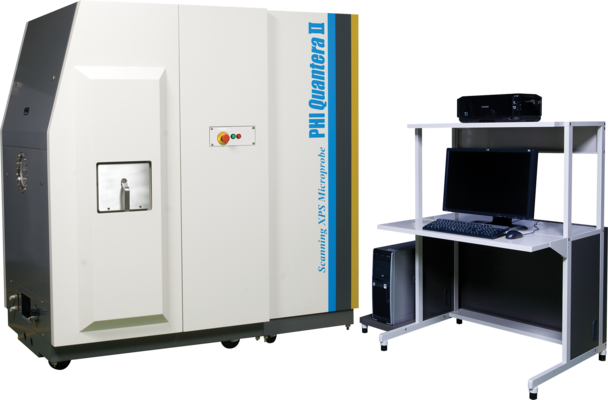
The core technology of the PHI Quantera II is PHI’s patented, monochromatic, micro-focused, scanning x-ray source which provides excellent large area and superior micro-area spectroscopy performance. Spectroscopy, depth profiling, and imaging can all be performed over the full range of x-ray beam sizes including the minimum x-ray beam size of less than 7.5 µm. In addition to superior XPS performance characteristics the PHI Quantera II provides two in situ sample parking stations which enables the automated analysis of all three sample platens in a single user defined analysis queue.
Intuitive Sample Navigation And Confident Analysis Area Identification
- The unique scanning X-ray microprobe allows SEM like navigation with point-and-click control
- X-ray induced secondary electron imaging (SXI) provides perfect correlation between imaged areas and spectroscopy
Optimized Depth Profiling
- Multiple ion gun options (monatomic Ar, C60, argon cluster GCIB) for a variety of organic, inorganic, and mixed materials
- Full 5-axis stage functionality including rotation/tilt and heating/cooling during sputtering
- Multipoint profiling within a single sputter crater for on/off defect analysis and precious samples
- Adjustable solid collection angle for improved angular resolution for Angle Resolved analysis with advanced software for high-throughput film structure analysis
Superior Micro-Area Analysis
- Highest small area sensitivity on the market
- <10 microns microprobe size in x and y
- Image registration for unattended automated micro-area analysis
Suite Of Specialized Solutions For Advanced Surface Analysis Needs
- Heating and Cooling in situ
- Electrochemical (biasing, polarization studies) experiments
- Glove Box Adapter
Micro-Focused Scanning X-ray Source
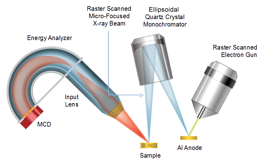
Unique Technology
- Micro-focused, raster scanned x-ray beam
- X-ray beam induced secondary electron imaging
- XPS images with spectra at each pixel for retrospective chemical analysis
- Point or multi-point spectroscopy
- Point or multi-point thin film analysis
Micro Area Spectroscopy
Unique Technology
- Micro-focused raster scanned x-ray beam
- Minimum beam size < 7.5 µm in diameter
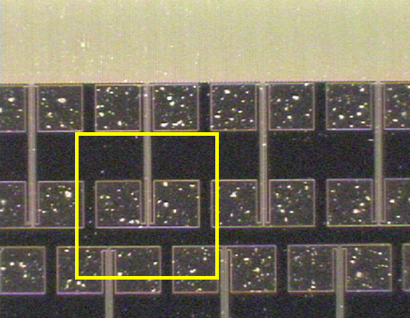
- Confidently locate small features for analysis using x-ray beam induced secondary electron images
- Highest small area XPS sensitivity
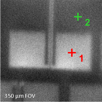
X-ray beam induced secondary electron image (SXI)
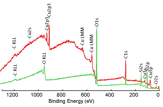
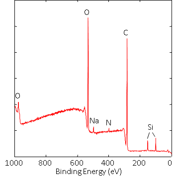
Spectra from selected areas obtained using a 20 µm diameter x-ray beam show Cu on the surface of the metal pads and SiO_2_ off of the pads.
Thin Film Analysis
Inorganic Thin Film Analysis
- 0-5 kV floating column ion gun
- Low voltage depth profiling for ultra-thin films
- Compucentric Zalar rotation
- Effective dual beam charge neutralization Bend in column to stop neutrals
- Micro-area depth profiling
- Multi-point depth profiling
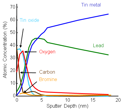
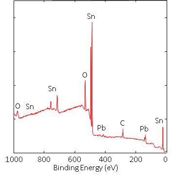
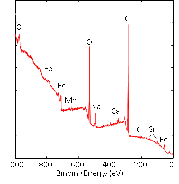
2 keV sputter depth profile of the surface species on a solder ball used for semiconductor packaging.
Organic Thin Film Analysis
- Optional Ar2500+ and C60 cluster source ion guns
- Mass filtered mass ion beam
- Sputters many polymer and organic materials without damaging their chemistry
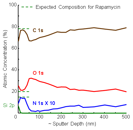
10 keV C_60_ depth profile of a 50/50 rapamycin and PLGA film showing segregation of the rapamycin to the surface of the coating.
Automated Analysis
High Throughput Automated Analysis
- Robust Auto-Z sample alignment
- Turnkey dual beam charge neutralization
- Move without concern from insulator to conductor in auto analysis sequences
- No special sample mounting or masking
Fully Automated Unattended Analysis
SXI Demo Video
SmartSoft-XPS
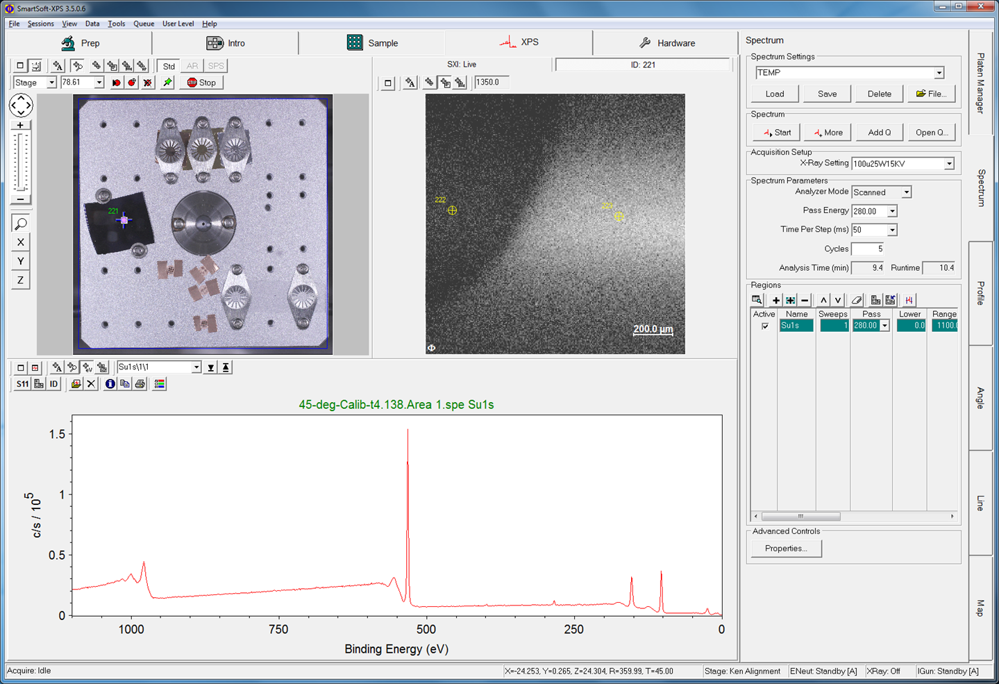
Whether you are a casual user or an expert, the work flow driven UI and enhanced feature set will increase your productivity.
- Intuitive single window user interface
- Session tabs guide you through the analysis process
- Integrated sample platen management
- Point and click analysis area definition on saved images
- User friendly queuing of multiple analysis tasks
- Multi-point analysis and sputter depth profiling within an imaged area
- Fully integrated control of optional accessories
MultiPak Data Reduction Software
Data Reduction for XPS and AES
PHI MultiPak is the most comprehensive data reduction and interpretation software package available for electron spectroscopy. The tasks of spectral peak identification, extracting chemical state information, quantification, and detection limit enhancement are addressed with an array of powerful and easy-to-use software tools for spectra, line scans, images and depth profiles. Microsoft Windows XP and Windows 7 compatible, MultiPak can be used on the instrument PC to process data in real time or on an off line PC for report generation.
Advanced Data Reduction Tools
- Auto peak identification
- XPS chemical state database
- XPS spectral deconvolution
- Quantitative analysis
- Non-linear least squares fitting
- Linear least squares fitting
- Target factor analysis
- Retrospective chemical imaging
- Batch mode data processing
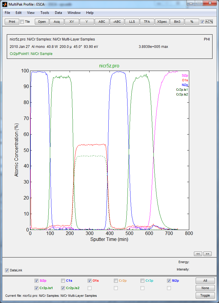
Chemical depth profile of a multi-layer Ni-Cr thin film structure showing the presence of Cr metal and Cr oxide layers.
Brochures
Application Notes
Learn how our instruments can help with your applications